
We are proud industry leaders in 3D Mammogram services, offering the highest level of breast radiology care with an unrivalled use of state-of-the-art mammogram technology, supported by our team of fully qualified, dedicated mammographers.

What is a Mammogram?
A 3D Mammogram, also known as Breast Tomosynthesis, uses high-powered computing to convert digital breast images into a stack of very thin layers or ‘slices’— building what is essentially a ‘three dimensional mammogram’. This gives our radiologist breast tissue detail in a way never possible before, to enable the breast tissue to be examined a millimeter at a time. 3D mammograms are standard for all Imaging Associates patients, inclusive of high definition synthetic 2D mammography, which minimises radiation dose.
At Imaging Associates, we have a team of specialised breast radiologists across our purpose built Mammography clinics, all housing state-of-the-art breast screening equipment, with adjoining ultrasound facilities.
This means that if you have a mammogram at an Imaging Associates radiology clinic in Melbourne or Wagga Wagga, there will be a breast radiologist (specialist) to review your mammogram straight after you have it, prepare the report, and be available for your referring doctor to talk to if required. This ensures the highest quality care across our clinics to help in the fight against breast cancer.
What Mammogram technology do we offer?
Our ongoing leadership in 3D mammography includes using the latest state-of-the-art technology including:
- Volpara – VolparaTM is software that can accurately and objectively measure mammographic breast density, and we provide the VolparaTM measurement of breast density on your mammogram report. Mammogram reports from other radiology clinics often do not give a clear subjective measure of breast density, let alone an objective measure such as a VolparaTM score. It is important to know your breast density, as having a high breast density for your age increases your risk of developing breast cancer.
- TransparaTM Artificial Intelligence – Improves the detection of breast cancer on mammograms by acting as an ‘extra pair of eyes’. We are the first site in Australia to offer this technology. Studies show TransparaTM improves both sensitivity and specificity in radiologist reporting of mammograms.
- Contrast Enhanced Mammography (CEM) – Mammography with an intravenous injection of contrast, which provides additional physiological information in the breast and further increases the detection rate of breast cancer over conventional mammography. Contrast mammograms can be performed in conjunction with 3D mammography.
How do I book a Mammogram near me?
Book a 3D Mammogram by completing our booking form or calling your nearest Imaging Associates radiology clinic now. Breast Imaging Services are available at the below Imaging Associates locations.
- Melbourne VIC – Carlton, Box Hill, Mitcham, Richmond
- Wagga Wagga NSW – Edward Street
Imaging Associates accepts all medical imaging referrals, even if written or printed on another radiology company’s request form.
Book an appointment
What do you need to know?
There is no preparation required for this examination. It’s recommended you wear a two-piece outfit and you will be advised not to wear talcum powder. Deodorant and perfume are fine.
Please advise us of any medication you are taking prior to your scan.
What do we need to know?
Please advise us if you are pregnant or think you might be pregnant, or if you are breastfeeding.
In this section
CT ScanLung Cancer ScreeningAbdomen CT ScansCT Pulmonary AngiogramCT Coronary AngiogramCT Calcium ScoreCT Lumbar SpineDental X-RayX-RayDEXA ScanFluoroscopyInterventional Breast ProceduresInterventional Radiology: Sports, Spinal and Pain Management3D MammogramMagnetic Resonance Imaging (MRI)Nuclear MedicineUltrasoundPregnancy UltrasoundWomen’s Imaging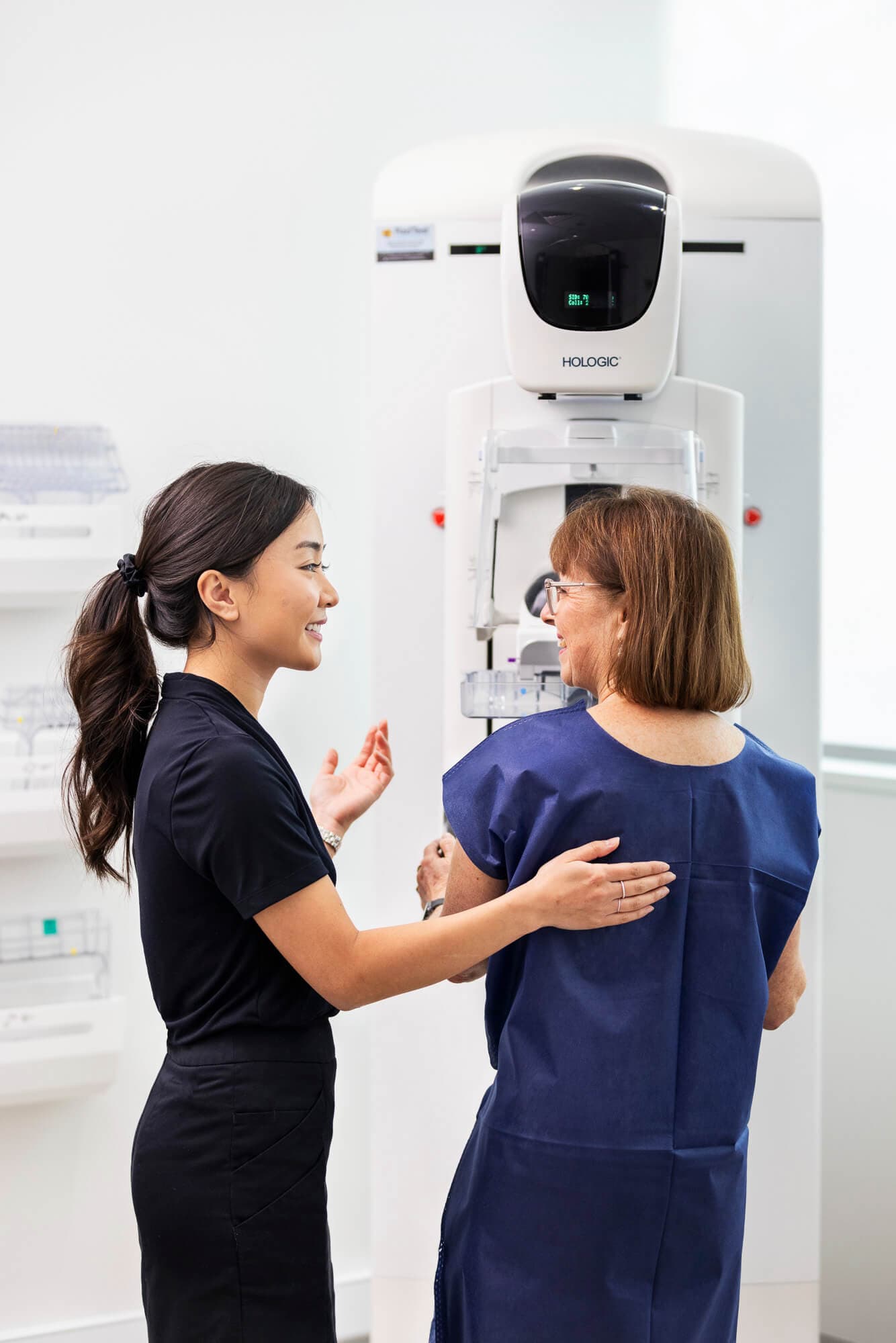
Book an appointment
Complete our online booking form to request an appointment and one of our staff will be in contact.
Book now

